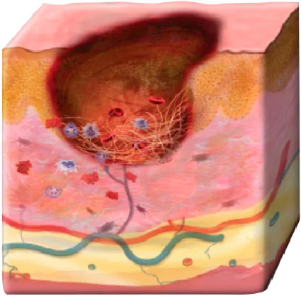
The challenges of healing stalled VLUs and DFUs.
A dysfunctional wound environment and comorbid conditions are major factors that may stall wounds. It’s imperative to heal VLUs and DFUs quickly because patients can experience serious complications, even death.1-5
Contributing factors to a dysfunctional wound environment
The stalled wound environment is inherently dysfunctional and is characterized by multiple factors that impede healing.
Biofilm is present in about 80% of chronic wounds, resulting in delayed healing by impairing the inflammatory response, formation of granulation tissue, and epithelialization.6,7
Repeated tissue injury stimulates the constant influx of immune cells, which results in a chronic release of pro-inflammatory cytokines that can damage nearby tissue.8,9
Fibrosis results from uncontrolled ECM production. This excessive deposition of collagens disrupts the proper tissue structure and function.10
Up-regulation of pro-inflammatory cytokines and chemokines inhibits normal progression of wound healing. This chronic inflammatory environment subjects various growth factors and cytokines to degradation and sequestration in the wound site, inhibiting their function.11
Chronic wound fibroblasts are unresponsive to stimulation by critical growth factors and show significantly decreased rates of ECM organization.12,13 Plus, keratinocytes have an inability to up-regulate chemokines and cytokines necessary for the resolution of chronic inflammation.14
In chronic wounds, MMP levels exceed that of their inhibitors, which leads to ECM degradation. This destruction of ECM prevents the wound from moving forward in the healing process and attracts more inflammatory cells, thus amplifying the inflammation cycle.8
Comorbid condition may negatively affect a patient’s ability to heal15,16
- Cardiovascular disease†
- Musculoskeletal conditions
- Obesity/overweight
- Neuropathy
- Peripheral vascular disease
- Foot deformities
- Cardiovascular disease†
- Musculoskeletal conditions
- Obesity/overweight
- Neuropathy
- Peripheral vascular disease
- Foot deformities
Why faster healing is critical for VLUs and DFUs
VLUs worsen over time, making them harder to heal
Percentage of wounds ≥1 year that did NOT heal17
44%
Size <10 cm2
78%
Size ≥10 cm2
VLUs significantly impact patients' quality of life
≤80%
Experience pain1,2
81%
Have impaired mobility18
68%
Report a negative
emotional impact18
2 million
Lost working days per year19
DFUs put patients at risk for serious complications, including osteomyelitis and amputation3,4
85%
Nontraumatic leg
amputations preceded
by DFU*20
15%
DFUs that result in lower
extremity amputation21
Every
5 minutes
A lower limb amputation occurs‡22
‡In patients with diabetes.Patients with DFUs or diabetes-related amputations have
5-year mortality
rates worse
than many common types of cancers23
Looking to overcome the healing challenges?
Discover how Apligraf® helps heal stalled VLUs and DFUs, or contact an Organogenesis Tissue Regeneration Specialist to see how Apligraf helps your VLU and DFU patients.
Contact usPlease refer to the Apligraf Package Insert for complete prescribing information.
REFERENCES:
- Gonzalez-Consuegra RV, et al. J Adv Nurs. 2011;67(5):926-944.
- Palfreyman S. Nurs Times. 2008;104(41):34-37.
- Giurato L, et al. World J Diabetes. 2017;8(4):135-142.
- Lipsky BA, et al. Diabetes Metab Res Rev. 2016;32(1 suppl):45-74.
- Moulik PK, et al. Diabetes Care. 2003;26(2):491-494.
- Malone M, et al. J Wound Care. 2017;26(1):20-25.
- Hurlow J, et al. Adv Wound Care. 2015;4(5):295-301.
- Frykberg RG, et al. Adv Wound Care. 2015;4(9):560-582.
- Seth AK, et al. J Surg Res. 2012;178(1):330-338.
- Giannandrea M, et al. Dis Model Mech. 2014;7(2):193-203.
- Barrientos S, et al. Wound Repair Regen. 2008;16(5):585-601.
- Cook H, et al. J Invest Dermatol. 2000;115(2):225-233.
- Kim BC, et al. J Cell Physiol. 2003;195(3):331-336.
- Wall IB, et al. J Invest Dermatol. 2008;128(10):2526-2540.
- Kelly M, et al. Int J Low Extrem Wounds. 2019;18(3):301-308.
- McEwen LN, et al. J Diabetes Complications. 2013;27(6):588-592.
- Margolis DJ, et al. Wound Repair Regen. 2004;12(2):163-168.
- Phillips T, et al. J Am Acad Dermatol. 1994;31(1):49-53.
- Sen CK, et al. Wound Repair Regen. 2009;17(6):763-771.
- Singh N, et al. JAMA. 2005;293(2):217-228.
- Snyder RJ, et al. Ostomy Wound Manage. 2009;55(11):28-38.
- Centers for Disease Control and Prevention. https://www.cdc.gov/diabetes/pdfs/data/statistics/national-diabetes-statistics-report.pdf. Accessed December 12, 2019.
- Armstrong DG, et al. Int Wound J. 2007;4(4):286-287.
